
Osteoarthritis is a degenerative joint disease that affects millions of people worldwide. It occurs when the protective cartilage that cushions the ends of bones in joints gradually wears away, leading to pain, stiffness, and swelling in the affected joint. Osteoarthritis can affect any joint in the body, but it is most commonly found in weight-bearing joints, such as the knees, hips, and spine.
One of the most common locations for osteoarthritis is the knee joint, and the distal medial femoral condyle is a frequent site for the development of osteoarthritis in the knee. The distal medial femoral condyle is the lower part of the thighbone that sits on the tibia, or shinbone. In osteoarthritis, the cartilage on the surface of the distal medial femoral condyle gradually breaks down, leading to inflammation and swelling in the surrounding tissues.
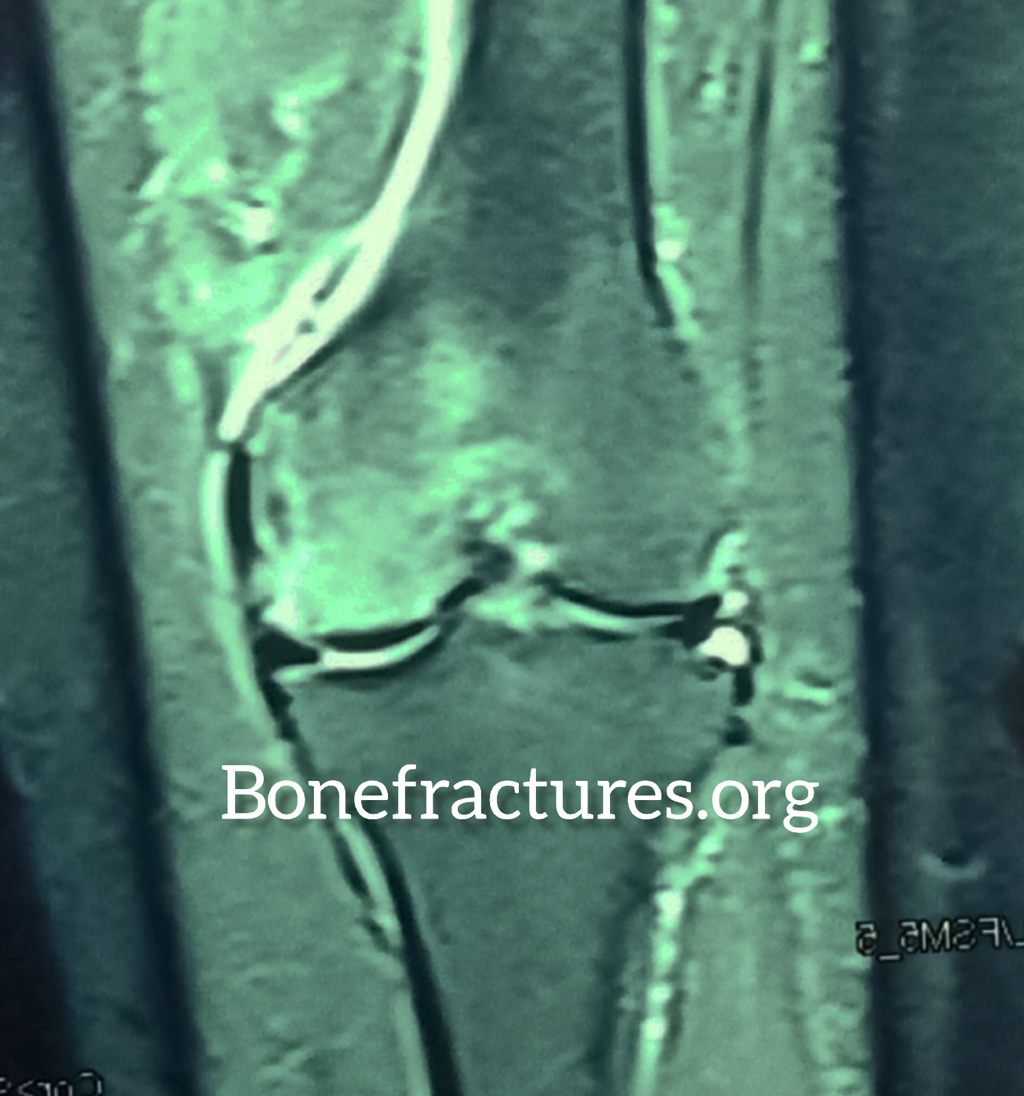
MRI is an excellent tool for diagnosing osteoarthritis because it can show the soft tissues, such as cartilage and ligaments, that cannot be seen on X-rays. The MRI sign of distal medial femoral condyle edema due to osteoarthritis is a specific finding that can be seen on an MRI scan.
When a person has osteoarthritis of the knee, an MRI scan of the affected joint will show increased fluid and swelling in the tissues surrounding the distal medial femoral condyle. This edema is a result of the inflammation caused by the breakdown of the cartilage in the joint. The edema appears as bright white areas on the MRI scan and is often accompanied by a loss of cartilage in the affected area.
In addition to the edema and loss of cartilage, an MRI scan of a knee affected by osteoarthritis may also show other changes, such as bone spurs, cysts, and thickening of the joint capsule. These changes can contribute to the pain, stiffness, and swelling that are characteristic of osteoarthritis.
In conclusion, the MRI sign of distal medial femoral condyle edema is a specific finding that can be seen in people with osteoarthritis of the knee. This edema is a result of the inflammation caused by the breakdown of the cartilage in the joint and appears as bright white areas on the MRI scan. An MRI scan is an excellent tool for diagnosing osteoarthritis because it can show the soft tissues, such as cartilage and ligaments, that cannot be seen on X-rays. If you are experiencing knee pain, stiffness, or swelling, speak to your doctor about whether an MRI scan may be appropriate for you.