


Plan your approach carefully: Before starting the surgery, it is important to carefully plan your approach. This includes reviewing imaging studies, assessing the degree of fracture displacement, and determining the optimal placement of the intramedullary nail.
Prepare the patient: Position the patient correctly on the operating table to allow access to the operative site, and provide appropriate anesthesia and pain management.
Use a reduction clamp: A reduction clamp is used to reduce the fracture and maintain the reduction during the fixation process. The clamp should be placed across the fracture site, and gentle traction should be applied to the leg while the clamp is tightened to achieve reduction.
Insert the intramedullary nail: Once reduction has been achieved, the intramedullary nail can be inserted through the fracture site. Care should be taken to avoid damaging the surrounding soft tissue.
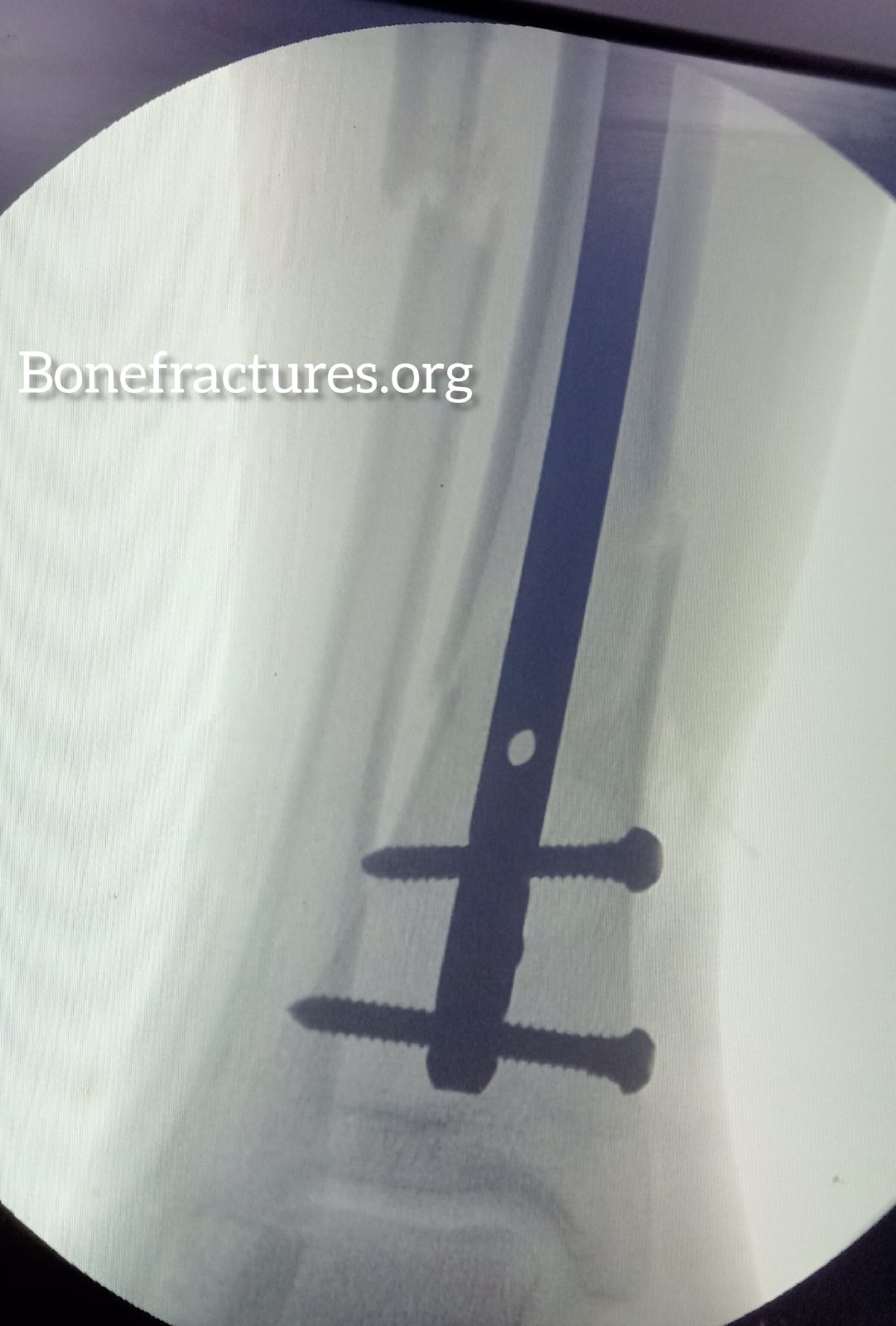
Confirm proper alignment: Confirm proper alignment of the fracture before inserting the nail fully, and after inserting the nail, using intraoperative imaging. The intramedullary nail should be positioned parallel to the joint line, with the proximal end of the nail just below the articular surface.
Complete fixation: Once proper alignment has been confirmed, complete the fixation by inserting interlocking screws at the proximal and distal ends of the nail to prevent rotation and ensure stability.
Close the incision: After fixation is complete, close the incision carefully and ensure that the skin edges are properly aligned.
Monitor the patient: Monitor the patient carefully postoperatively for signs of infection, neurovascular compromise, or other complications. Provide appropriate pain management and rehabilitation as needed.